BIOL 1021 DU Mammalian & Epithelial Tissue Epidermis & Epithelium Cells Lab Report
Question Description
Mammalian Tissues
Background
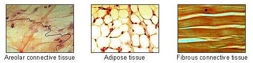
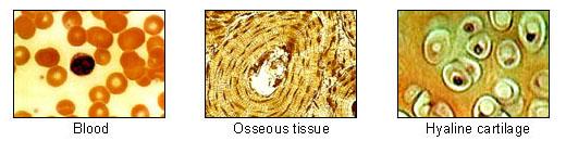
Figure 1. Cells Have Unique Appearances
A human body begins as a single cell. As the individual develops, many different cell types differentiate from this single cell. All of your cells have the same genome, but many types of cells look very different from each other (Figure 1).
Cells and Tissues
Tissue refers to a collection of cells that perform a single function together. Combinations of various tissues come together to form organs within an organism. The study of these various tissues is called histology.
There are four basic categories of animal tissues:
- Epithelial tissue
- Connective tissue
- Muscle tissue
- Nervous tissue
Each tissue type looks distinct from the others and serves a unique purpose.
Epithelial Tissue
Epithelial tissue covers the surfaces of organs and forms the organisms outer surface. Structurally, epithelial cells are tightly bound together with little space between them. This structure leads to function. The tightly bound epithelial cells form epithelial tissue that is a barrier that impedes or allows appropriate substances to pass through.
Epithelial tissue is classified based on the grouping and shape of the cells that make up the tissue (Figure 2).
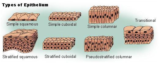
Figure 2. Epithelial Tissue Types
The name of most types of epithelial tissue can be broken into two terms. The first term describes the epithelial cell group:
- Simple: a single layer of epithelial cells
- Stratified: multiple layers of epithelial cells, one on top of the other
The second term describes the basic epithelial cell shapes:
- Squamous: flattened
- Cuboidal: cube-like
- Columnar: tall and thin
Connective Tissue
Connective tissue (Figure 3) fills spaces, stores fat, transports materials, and binds, supports, and protects body parts. Although different connective tissues are seemingly very different, cells are all spaced far apart and have a substance between the cells. This substance, called the extracellular matrix, is composed of materials that range from elastic fibers to fibers made rigid with calcium salts to gelatinous protein solutions to fluid blood plasma.

Figure 3. Examples of Connective Tissue Types (click here for larger view )
There are several types of connective tissue:
- Loose connective tissue is composed of thick collagen fibers, thin elastin fibers, and the fibroblast cells that secrete the proteins composing these two types of fibers. Adipose, reticular, and areolar are the three kinds of loose connective tissue. Adipose cells are so full of lipid storage that the nuclei are pressed against the membrane. Cells in areolar and reticular tissues are surrounded by large amounts of extracellular matrix full of elastic fibers.
- Dense connective tissue, similar to loose connective tissue, is made up of collagen fibers, elastin fibers, and fibroblast cells. However, unlike loose connective tissue, the fibers of the dense connective tissue are packed so closely together that there are few open spaces. Dense connective tissue is strong and flexible. It forms strong, rope-like structures, such as tendons and ligaments, which connect bones and muscles. Dense connective tissue can also be arranged in sheets, such as in the dermis in the skin. It provides the skin with elasticity and strength, so the skin does not tear.
- Cartilage is the smooth material found in joints where the ends of bones meet and move against each other, such as in the knee, hip, elbow, and shoulder. It is also found in the nose and the walls of the respiratory passages. The matrix of cartilage is chondrin. Chondrin is a gelatinous mixture of proteins and carbohydrates secreted by cells called chondrocytes. Chondrocytes are loosely spread throughout the matrix of cartilage and inside cavities called lacunae (singular: lacuna), found singularly or in small groups.
- Bone is composed of a matrix that is hard, dense, and composed of calcium salts deposited on loose collagen fibers. Because the matrix is hard, cells are not found loose within the matrix but in lacunae, or cavities embedded with the cells. The cells in the lacunae that make the matrix are called osteocytes. In compact bone, bone tissue grows in concentric rings around a central canal called a Haversian canal. Blood vessels and nerves pass through these canals. This entire structure of rings with a central canal is called an osteon. When grouped together, many osteons form a compact bone.
- Blood is considered a type of connective tissue. Its matrix consists of fluid-like plasma made up of proteins, glucose, clotting factors, mineral ions, hormones, and carbon dioxide dissolved in water. The cells suspended in the plasma are erythrocytes (red blood cells), which are shaped like small disks pinched in the center. They deliver oxygen to all the organs and remove carbon dioxide. Leukocytes (white blood cells) are involved in the bodys immune response to disease and foreign invasion.
Muscle Tissue
Muscle tissue has cells arranged in long, threadlike fibers, some of which can contract when an electrical impulse is applied. Muscles are the bodys motors, functioning to produce movement. There are three types of muscle tissue (Figure 4).
- Skeletal muscle is found in the muscles attached to bones. It is composed of long, branching fibers with striations, or stripes of alternating color, perpendicular to the long axis of the fibers and multinucleated cells, or single cells containing multiple nuclei. It is the only muscle tissue under voluntary control.
- Cardiac muscle is found only in the heart. It is striated and arranged in long fibers, like skeletal muscle. Unlike skeletal muscle, its cells are not multinucleated. Notably, cardiac muscle has intercalated disks, which are thick lines running the width of the muscle fibers like dividers. These disks speed up the flow of electrical current throughout the heart, which allows the entire organ to contract in a smooth motion on every heartbeat.
- Smooth muscle, unlike the other two muscle tissues, is nonstriated. Smooth muscle is associated with various organs, such as the stomach and intestines.
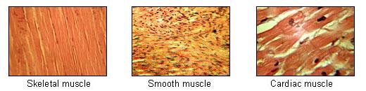
Figure 4. Muscle Tissue Types
Nervous Tissue
Nervous tissue is found in the brain, spinal cord, and nerves. It is made of interconnected cells called nerve cells, or neurons, that transmit both information and commands throughout the organism. Many tiny cells called glial cells surround the neurons and provide physical and nutritional support.
Nerve cells have a unique form with a large cell body, called the soma, and many arm-like extensions, called dendrites, radiating from it (Figure 5). Each nerve cell has:
- A singular long axon that carries electrical impulses away from the nerve cell
- Many, shorter dendrites that connect to the axons of other nerve cells and receive electrical impulses

Figure 5. Nerve Cells (click here for larger view )
Complex Organ Structures
Groups of tissues together make up organs. Each type of organ has a unique structure, which leads to a unique function. Two examples of organs are the skin and blood vessels.
Skin
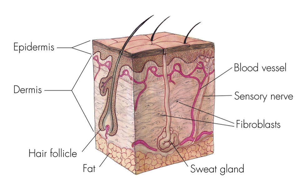
Skin covers and protects the entire body, but it is not impenetrable. In fact, hairs and secretions emerge from its surface, and skin is very pliable. That pliability allows the skin to maintain its structural integrity while moving and when encountering external surfaces. Secretions help maintain this suppleness. The skin is made up of three tissue layers:
- The outermost layer of the skin, or epidermis, is composed of dead epithelial cells. Epidermal cells are flattened, misshapen, and filled with a protein called keratin. Keratin is the material that fingernails are made of. It is tough and water resistant. Epidermal cells are constantly being worn down and falling off and must, therefore, be constantly replenished.
- The layer below the epidermis is the dermis. Here you will find specialized epithelial cells called glands. The oil gland, or sebaceous gland, is an alveolar gland that empties secretions that lubricate the hair into the channel through which the hair follicle exits the surface of the skin. Alveolar means that is shaped like a cluster of sacs, similar to a bunch of grapes.
- Beneath the dermis is the hypodermis where there are many round, seemingly empty, fat cells. However, if you look closely you will see some with their nuclei pressed right up against the cell membrane. Fat cells are a kind of loose connective tissue called adipose tissue.
Blood Vessels
Arteries and veins are two types of blood vessels that transport blood to and from body tissues. Arteries control the flow of blood by dilating and contracting, whereas veins use valves to prevent backflow. There are several structural differences between the two types of blood vessels that allow for the different ways the blood vessels function:
- Arteries have thicker walls than veins do.
- Arteries have a thicker layer of smooth muscle than veins do.
- The lumen (inner diameter) of arteries is generally smaller than that of veins of comparable sizes.
The tissues that make up both the arterial and venous walls are divided into three concentric layers, called tunica (plural: tunicae). From the inside outward, they are:
- Tunica intima: a single layer of epithelial cells with an elastic membrane
- Tunica media: circular smooth muscle
- Tunica externa or Tunica adventitia: connective tissue surrounding the blood vessel and an outer elastic membrane
About This Lab
In this lab, you will learn to recognize the different tissue types and see how a collection of tissues form organs.
Experiments
Open the simulation by clicking on the virtual lab icon below. The simulation will launch in a new window.
You may need to move or resize the window in order to view both the Procedure and the simulation at the same time.
Follow the instructions in the Procedure to complete each part of the simulation. When instructed to record your observations, record data, or complete calculations, record them for your own records in order to use them later to complete the post-lab assignment.

Procedures
Experiment 1: Epithelial Tissue
- View the two images of epithelial tissues below.
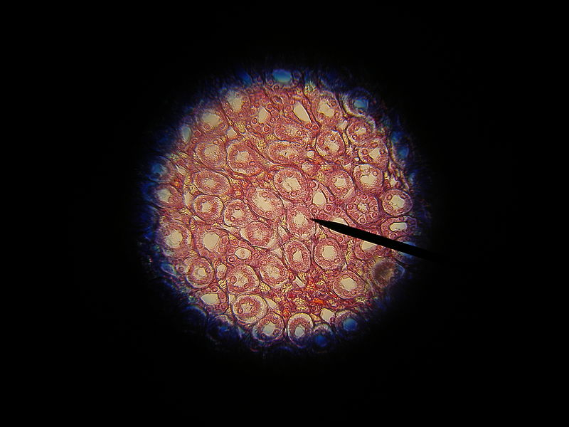
Epithelial Tissue from Kidney
Epithelial Tissue from Dermis and Epidermis
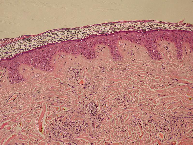
- For each image, find the epithelial cells. Record a description of their shape and structure to reference later. Note that the individual cells are very small. Refer to the information provided in the Background in your Hayden-McNeil course site to find written and visual descriptions of the different types of epithelial tissues.
Experiment 2: Connective Tissue
Part 1: Loose Connective Tissue
- View the diagram of areolar connective tissue.
Areolar Connective Tissue

- Record descriptions of the three components of loose connective tissue to reference later.
- Collagen fibers
- Elastin fibers
- Fibroblast cells
Use the labels on the image to determine any implied components of the tissue.
Part 2: Dense Connective Tissue
- View the diagram of dense connective tissue. The image shows labeled collagen fibers and the nuclei of fibrocytes (fibroblasts). Though elastin fibers are found in dense connective tissue, this component is not labeled in the image.
Dense Connective Tissue
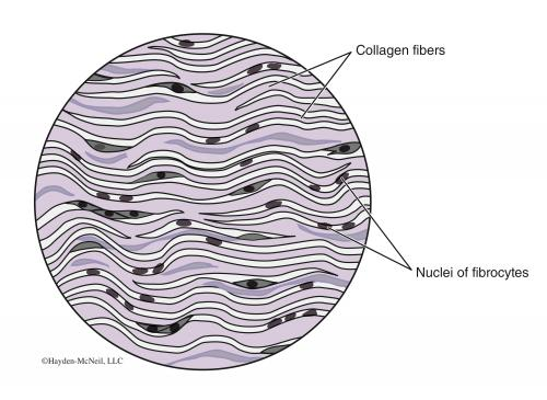
- How does dense connective tissue appear? How does it differ from loose connective tissue? Record your answers to reference later.
- Compare the following components that are labeled in both the loose connective tissue and dense connective tissue images.
- Collagen fibers
- Fibroblast cells
Record your observations to reference later.
Part 3: Cartilage
- View the diagram of elastic cartilage.
Elastic Cartilage
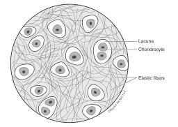
- The gel matrix is not labeled on the diagram. Note that the gray material in which the elastic fibers appear is the gel matrix.
- Record your descriptions of the following components of cartilage to reference later.
- Chondrocytes
- Lacunae
- Gel matrix
Part 4: Bone
- View the diagram of bone tissue.
Bone Tissue Diagram
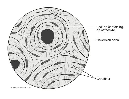
- Familiarize yourself with the appearance of the following components.
- Haversian canal
- Lacuna and osteocyte
- An osteon is made up of a Haversian canal and the structure of rings around it. Find an osteon in the diagram.
- View the image of bone tissue.
Bone Tissue Image
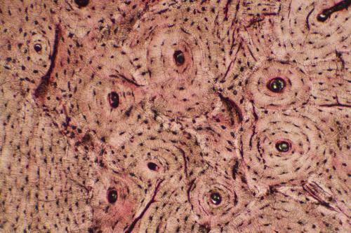
- Observe the following components in the image above.
- Haversian canal
- Lacuna and osteocyte
- Osteon
- Record your observations about the bone tissue to reference later.
Experiment 3: Muscle Tissue
- View the skeletal muscle tissue diagram.
Muscle Tissue Diagram

- Familiarize yourself with the following components. Use the labels on the diagram to determine any implied components of the tissue.
- Muscle cells and fibers
- Nuclei
- Striations along a fiber
- Take the microscope from the Instruments shelf and place it onto the workbench. Click here for a short video on using the virtual microscope.
- Take the skeletal muscle slide from the Containers shelf and place it on the microscope stage.
- Explore the slide until you find one or more views that show each of the components identified in the diagram above.
- Label the following components on one or more views of the microscope slide, using the diagram above as reference.
- Nuclei
- Striations along a fiber
- Take screenshots of each of your annotated images in order to upload and submit them later.
- Using your draw overlay tool, draw a line in the direction in which the muscle fibers are aligned.
- Save a screenshot of the annotated image.
- When you are finished, drag the slide back to the Containers shelf.
Experiment 4: Nervous Tissue
- View the neuron diagram.
Neuron Diagram
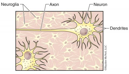
- Familiarize yourself with the following components.
- Dendrites
- Axon
- Neuroglia (glial cells)
- Take the nervous tissue slide from the Containers shelf and place it on the microscope stage.
- Label the following components on one or more views of the microscope slide, using the diagram above as reference.
- Dendrites
- Axon
- Neuroglia (glial cells)
- Take screenshots of each of your annotated images in order to upload and submit them later.
- When you are finished, drag the slide back to the Containers shelf.
Experiment 5: Complex Organ Structures
Part 1: Skin
- View the diagram of skin in cross section below. Use it as a reference in this Part, as needed.
Skin Cross Section Diagram
- Take the skin cross section slide from the Containers shelf and place it on the microscope stage.
- Observe the epidermis. You may need to use the y-axis stage adjust knob to locate it. Increase the magnification of the microscope so that you have a close-up view of the epidermal layer. Note: In this slide, the epidermis stains much darker than the dermis underneath it.
- Use the annotation tools to label the epidermis. Take a screenshot of the annotated view in order to upload and submit it later.
- Record your observations about the epidermis to reference later. For example, how thick is the epidermis in number of cells? Its thickness is not uniform, so give a range from minimum to maximum. Can you tell what type of epithelial tissue makes up the epidermis? Why or why not?
- Reduce the magnification of the microscope so that you are viewing most of the cross section, from the epidermis to the bottom of the hair root.
- Observe the dermis.
- Annotate the slide view in the following ways.
- Circle and label a hair follicle
- Circle and label a group of fat cells
- Label the nucleus of a fat cell
Take a screenshot of the annotated view. You may need to use multiple views and take multiple screenshots.
- When you are finished, drag the slide back to the Containers shelf.
- Take the outer skin surface slide from the Containers shelf and place it on the microscope stage. This slide contains a slice of skin mounted flat for viewing from the top.
- How do these cells differ from the epithelial cells found beneath this layer, which you viewed in the previous slide? Record your observations to reference later. Include answers to the questions below.
- What is different about the shape of these cells?
- Do you see nuclei or any other internal cell structures?
- When you are finished, drag the slide back to the Containers shelf.
Part 2: Arteries and Veins
- View the artery and vein diagram. Use it as a reference in this Part, as needed.
Artery and Vein Diagram

- Take the artery and vein slide from the Containers shelf and place it on the microscope stage. This slide shows one artery and one vein in cross section, surrounded by connective tissue.
- At low magnification, identify and label the artery and the vein based on the anatomical differences between them.
- Save a screenshot of the annotated slide in order to upload and submit it later.
- Increase the magnification and focus on the artery.
- Identify and label the following components.
- Tunica intima
- Tunica media
- Tunica adventitia
- Save a screenshot of the annotated slide.
- Focus on the vein.
- Identify and label the following components.
- Tunica intima
- Tunica media
- Tunica adventitia
- Save a screenshot of the annotated slide.
- Clear the bench of all slides and instruments by dragging them back to the shelves, then return to your course page to complete any assignment for this lab.


